Structure and function of the eyeSpec 2.91
In short
The eye is a sense organ that detects light and sends impulses to the brain along the optic nerve. The cornea and lens refract light to focus it on the retina, which contains rod cells, sensitive to dim light, and cone cells, which detect colour. The iris controls the size of the pupil and so how much light enters.
The eye is a sense organ and acts as a receptor. It detects light and sends impulses to the brain.
| Part | Function |
|---|---|
| Sclera | Tough white outer layer that protects the eye |
| Cornea | Transparent front surface. It refracts (bends) light as it enters the eye |
| Iris | Ring of muscle that changes the size of the pupil, so controls how much light enters the eye |
| Pupil | The hole in the middle of the iris that lets light through |
| Lens | Refracts light to focus it on the retina |
| Ciliary muscles | Ring of muscle around the lens. Contracts or relaxes to change the shape of the lens |
| Suspensory ligaments | Hold the lens in place and connect it to the ciliary muscles |
| Retina | Contains the light receptor cells (rods and cones) |
| Fovea | Part of the retina with the most cones. Gives the sharpest, most detailed vision |
| Blind spot | Where the optic nerve leaves the eye. It has no receptor cells, so no light is detected there |
| Optic nerve | Carries impulses from the retina to the brain |
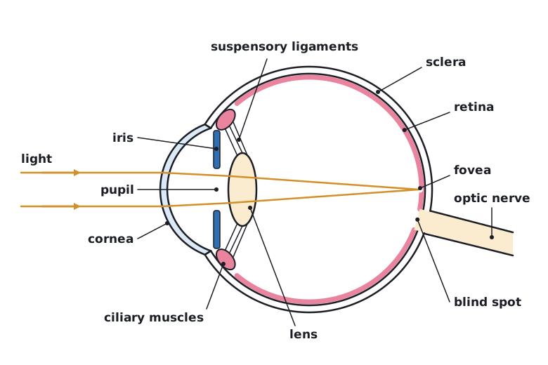 (opens full size in a new tab)
Rods and cones
The retina has two types of receptor cell. Rod cells are sensitive to dim light but do not detect colour. Cone cells detect colour and work in bright light.
Give each part with its job and a reason, for example ‘the cornea refracts light’, not just ‘the cornea is at the front’.
Written and checked against the Edexcel IGCSE Biology (4BI1) specification · Updated October 2026